Description
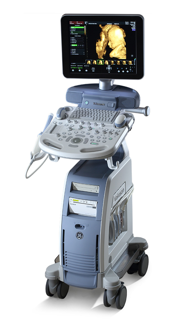
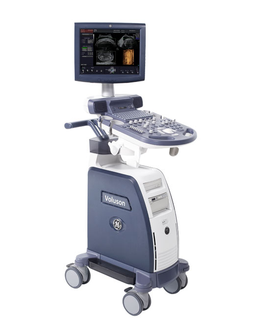
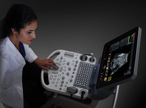
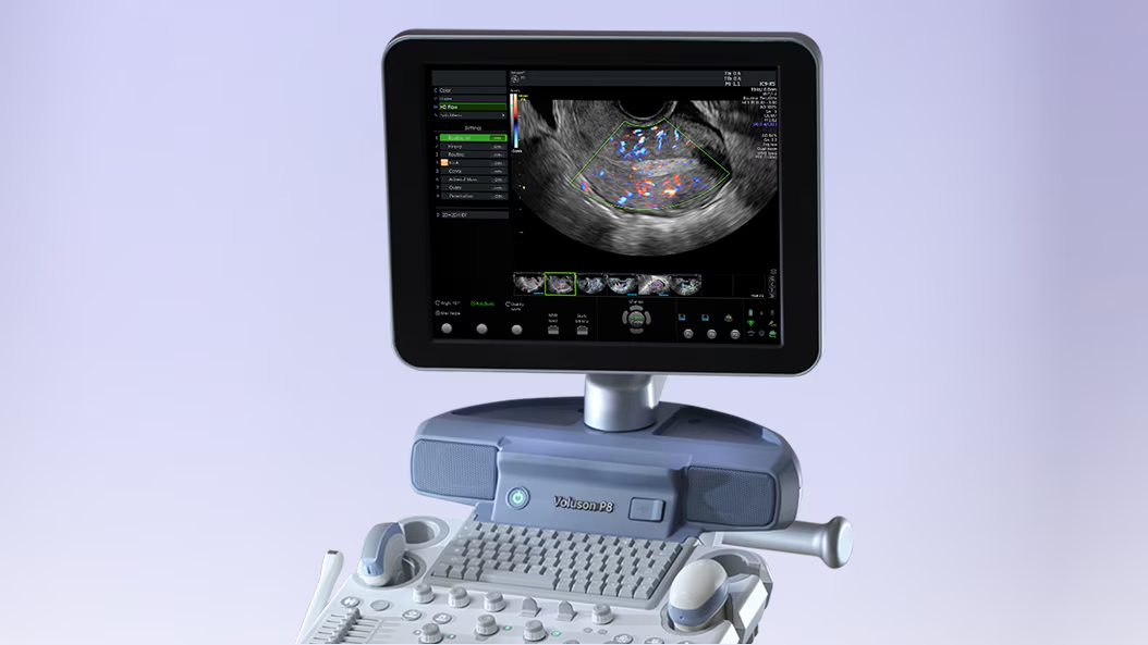
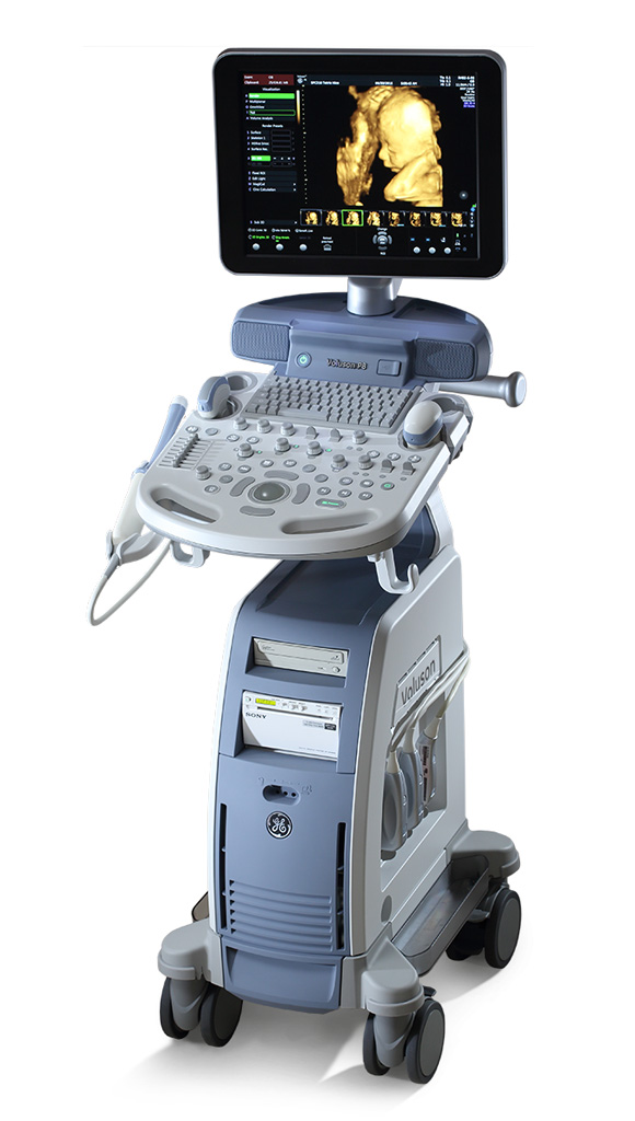
Voluson™ P8 ultrasound scanner (ultrasound) – a leading model in the Voluson Performance Series of ultrasound scanners for women’s health.
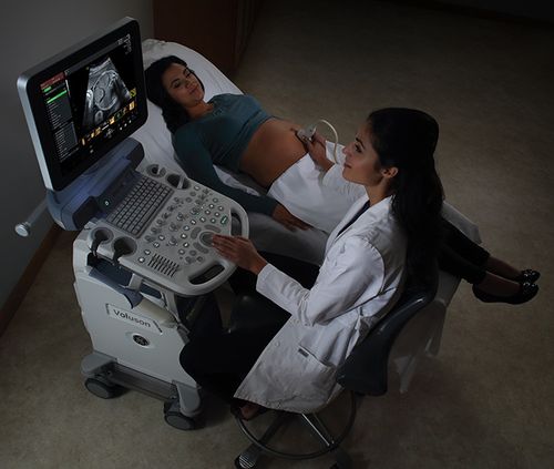
Visualisation is becoming easier. Ultrasound is an important tool to ensure high-quality women’s health.
1. The Voluson™ P8 delivers superb image quality and is easy to use and a streamlined workflow in a sleek and lightweight body.
2. Ideal for expanding your practice and handling heavy loads, The P8 delivers reliable, effective diagnostics across a range of clinical applications, including obstetrics, gynaecology, assisted reproductive technology and childbirth.
3. Its high performance, operational availability and exceptional reliability make the P8 a cost-effective way to join the Voluson family.
The Voluson P8 ultrasound system offers:
The ability to assess the coronary scan plane during 3D examinations for a detailed evaluation of anatomical structures.
Produce stunning 2D images without any additional settings.
A simple interface and user-friendly system design.
Assistive technologies are available.
High-quality visualisation with no extra effort
Excellent 2D images – with optimal settings for maximum clarity and detail.
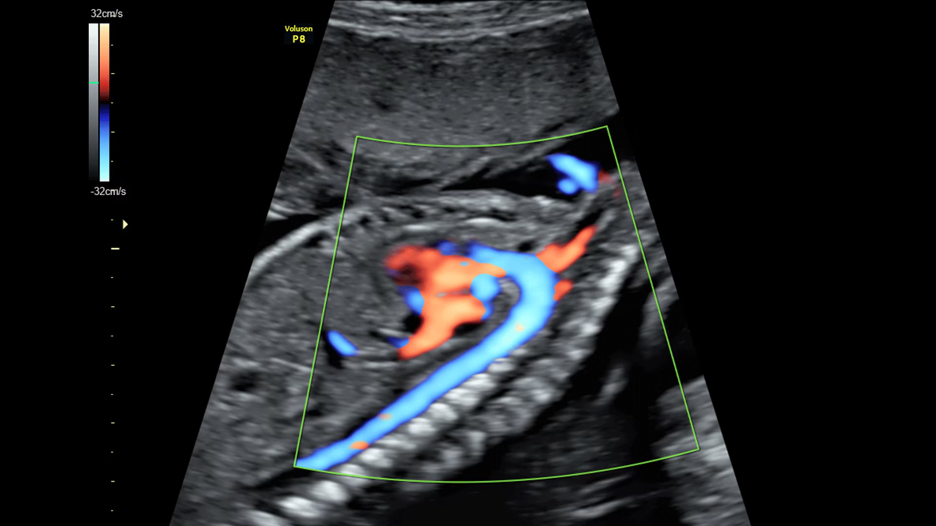
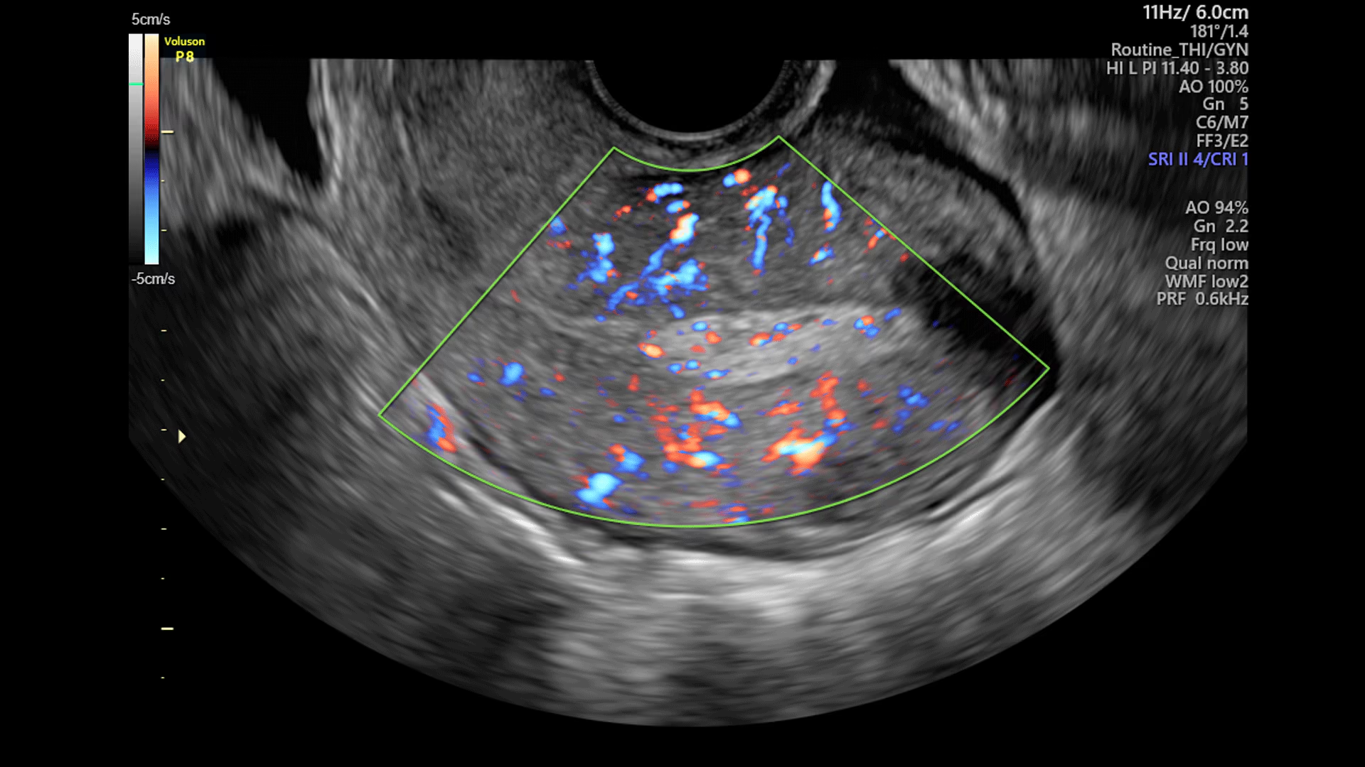
Advanced colour Doppler mapping – for a more detailed examination of the anatomy and functional status of the vascular system.
Excellent imaging at depth without any loss of quality – making it easier to examine complex patients.
One-touch image optimisation – for fast and effective visualisation.
Ease of obtaining 3D images
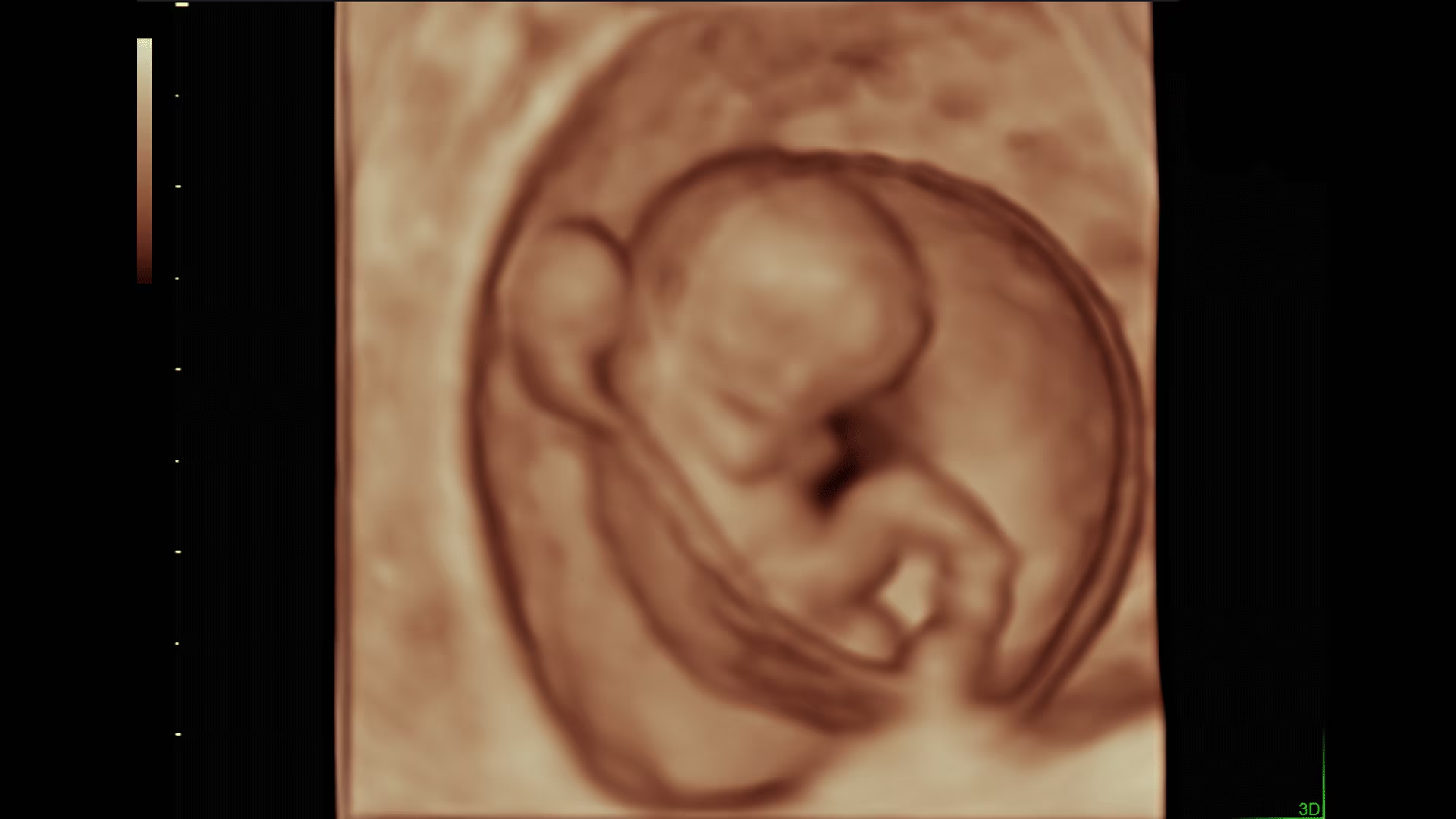
3D/4D imaging allows you to examine additional anatomical planes, boosting your confidence in making a diagnosis.
Visualisation of areas that cannot be examined using 2D scanning, for a more accurate diagnosis.
High productivity, thanks to the ability to analyse 3D data both during and after the study has been completed.
Viewing and processing 3D images in different planes or using different scanning modes.
The 17-inch LCD monitor provides high-quality display in all lighting conditions. It can be moved independently of the console, making it easy to adjust and convenient to use.
The built-in keyboard provides quick access to frequently used functions, helping to reduce the time spent searching for them and making the system more user-friendly.
Where it is used
- Perinatal centres | Maternity hospitals
- Diagnosis of superficial organs | Diagnosis of the breasts
- Inpatient Care | Clinical Diagnostics | Medical Laboratories
- Obstetrics and Gynaecology | Reproductive Medicine | Neonatology
- Gynaecology | Urology
Exceptional imaging performance starts with the Voluson Core Architecture.
The migration of expert technologies expands the capabilities of the Voluson™ P8 ultrasound scanner to improve image quality, frame rate and depth of penetration.
Excellent imaging for a wide range of patient types and examinations.
Simply pick up the sensor to get the information you need without any lengthy set-up.
- ■ 3D/4D allows for the acquisition of cross-sections that are not available in 2D, thereby increasing clinical confidence;
- ■ High-sensitivity colour Doppler enables the assessment of the foetus’s anatomy and well-being;
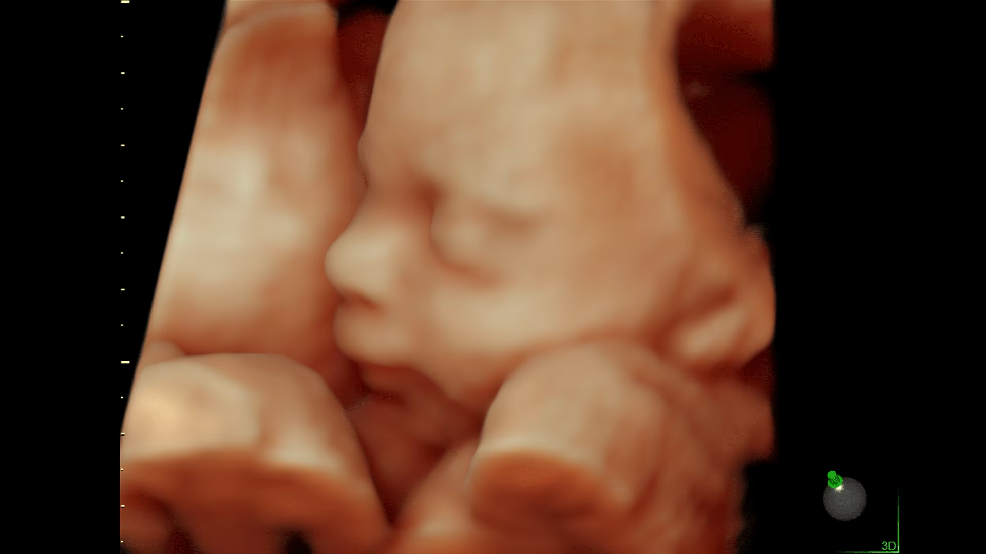
HDlive™️ Discover a unique clinical perspective (movable virtual light source).
The availability of additional information can help to enrich the dialogue between the clinician and the patient and contribute to a more comprehensive understanding of the examination results.
Gain more detailed information with less effort, thanks to the technology built into the Voluson™ P8:
- HD-Flow – improved visualisation of small vessels: thanks to clear imaging in 2D mode and increased sensitivity compared with conventional colour Doppler, making it ideal for assessing placental blood flow, the foetus’s small vessels and gynaecological examinations;
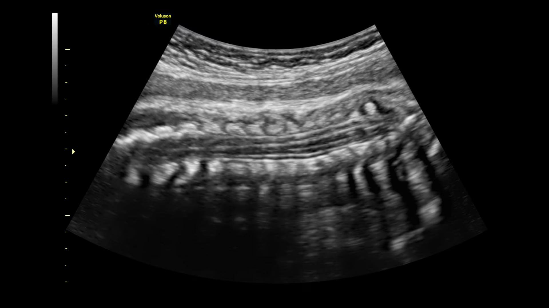
- OmniView – Easily visualise irregularly shaped structures, such as the uterus or the foetal spine. Simply draw a line through the structure to obtain views that are not available in 2D mode;
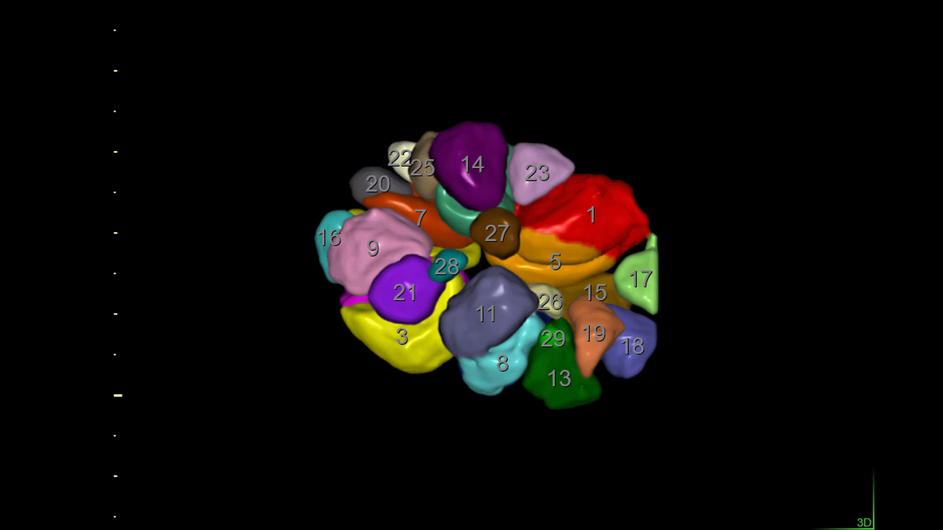
- SonoAVC™️follicle – Determines the number and calculates the dimensions and volume of hypoechoic structures;
- SonoFHR – Places the calipers in M or PW mode to automate the calculation of the foetal heart rate;
- SonoL&D – Measures and documents the descent of the foetal head, providing objective data for reporting and communication with the patient.
Discover 3D printing
Export files directly from the system to create 3D scans instantly, saving time and reducing complexity compared to using external post-processing software.
The Voluson™ P8 is lightweight and manoeuvrable
- ■ 17-inch high-resolution LED monitor
- ■ Battery (sleep mode – quick start)
- ■ Automatic image optimisation
- ■ Expansion of the scanning sector
- ■ CrossXBeam™ (CRI) multi-vector scanning
- ■ Multi-view 3D rendering
- ■ Anatomical M-mode
- ■ 4D
- ■ HDliveTM
- ■ Saving in a format suitable for 3D printing
Automated biometric measurements
- ■ SonoAVCfollicle automatic follicle measurement
- ■ Automatic measurement of foetal head descent during labour with SonoL&D
- ■ Automatic measurement of the collar zone and the fourth ventricle using SonoNT / SonoIT
- ■ Automatic measurement of foetal biometric data
- ■ Automatic measurement of the foetal heart rate
- ■ Automatic real-time alignment of the reconstruction plane with SonoRenderlive
Editing minutes.
A built-in archive with a preview function.
Educational videos.
-
Technical specifications
-
Core System Architecture Platform
-
17-inch high-resolution LED monitor
-
Sensor ports 3 active sensor ports
-
Battery: Sleep Mode – Quick Start
-
Automatic image optimisation Yes
-
Expanding the scanning area Yes
-
Educational videos Yes
-
-
Modes and features
-
Radiantflow Not available
-
HD-Flow high-sensitivity colour mapping Yes
-
B-Flow™ – non-Doppler blood flow mapping None
-
Multi-view 3D rendering Yes
-
STIC technology None
-
Anatomical M-mode Yes
-
Real-Time technologies Real-Time 4D
-
Visualisation modes (Hdlive, Silhouette) Hdlive – realistic rendering of 3D models
-
Uterine Trace – coronal section of the uterus None
-
Save in a format suitable for 3D printing Yes
-
Advanced VCI with OmniView – thick section, reconstruction along an arbitrary line None
-
Elastography (including analysis) Not available
-
Coded contrast harmonica None
-
Scan Assistant Not available
-
Editing minutes Editing minutes
-
Built-in archive with preview function Yes
-
SlowflowHD and Slowflow3D Not available
-
Radiant, UltraHD and Augment Not available
-
Advanced Fetal Echo Not available
-
fetalHQ None
-
AI-driven survey management None
-
AI and automated technologies None
-
XDclear™ Probes None
-
High-frequency endocavity sensors None
-
-
Automated biometric technologies
-
SonoAVCfollicle automatic follicle measurement Yes
-
SonoAVCantral 2.0 automatic follicle measurement Yes
-
Automatic measurement of foetal head descent during labour SonoL&D Yes
-
Automatic 3D measurement of the foetal head’s descent during labour Yes
-
Automatic measurement of the collar zone and the fourth ventricle in SonoNT / SonoIT Yes
-
Automatic generation of standard foetal heart sections using SonoVCADheart None
-
SonoCNS – automatic acquisition and measurement of foetal brain structures None
-
Automatic measurement of foetal biometric data Yes
-
Automatic measurement of the foetal heart rate Yes
-
SonoRenderlive’s automatic real-time alignment of the reconstruction plane Yes
-
-
Additional features
-
Changing the scanning angle for the BetaView TV sensor Yes
-
Patient database Yes
-
Image archive on the hard drive Yes
-
3D/4D data compression Yes
-
Real-time Doppler calculations Yes
-
Multigestational Calculations Yes
-
IOTA (International Ovarian Tumour Analysis): Yes
-
LR2, Simple Rules and the ADNEX Model Yes
-
IETA (International Endometrial Tumour Analysis) Report Yes
-
IDEA (International Deep Endometriosis Analysis) Report Yes
-
Tool for assessing sensor performance: Yes
-
-
Measurement and calculation packages, including reports and statements
-
OB Yes
-
GYN Yes
-
Abdominal Yes
-
Paediatrics Yes
-
Small Parts Yes
-
Musculoskeletal (MSK) Yes
-
Vascular Yes
-
Cephalic Yes
-
Transrectal Yes
-
Cardio Yes
-
Breast Yes
-
The price of the equipment is included:
-
Installation and configuration
-
Training in the use of equipment
-
24-month official warranty
-
Service and commissioning
-
Fast delivery
VAT